Article titled “Impact of TiO2 nanomaterials with different morphologies and their calcium phosphate composites on hemostasis and immunocompatibility” was accepted for publishing in Bulletin of Materials Science

The abstract of the paper is here:

The increasing use of TiO2 nanomaterials (TiNMs) in bone tissue engineering raises
concerns about their biocompatibility. To date, majority of biological testing was focused on the cytotoxicity of TiO2 nanoparticles and their composites. However, evaluation of immunocompatibility and effects on hemostasis is crucial for successful clinical applications of bone regeneration materials. Such data are scarce for TiNMs.  
To fill this gap, this study aimed to investigate impact of TiNMs with different
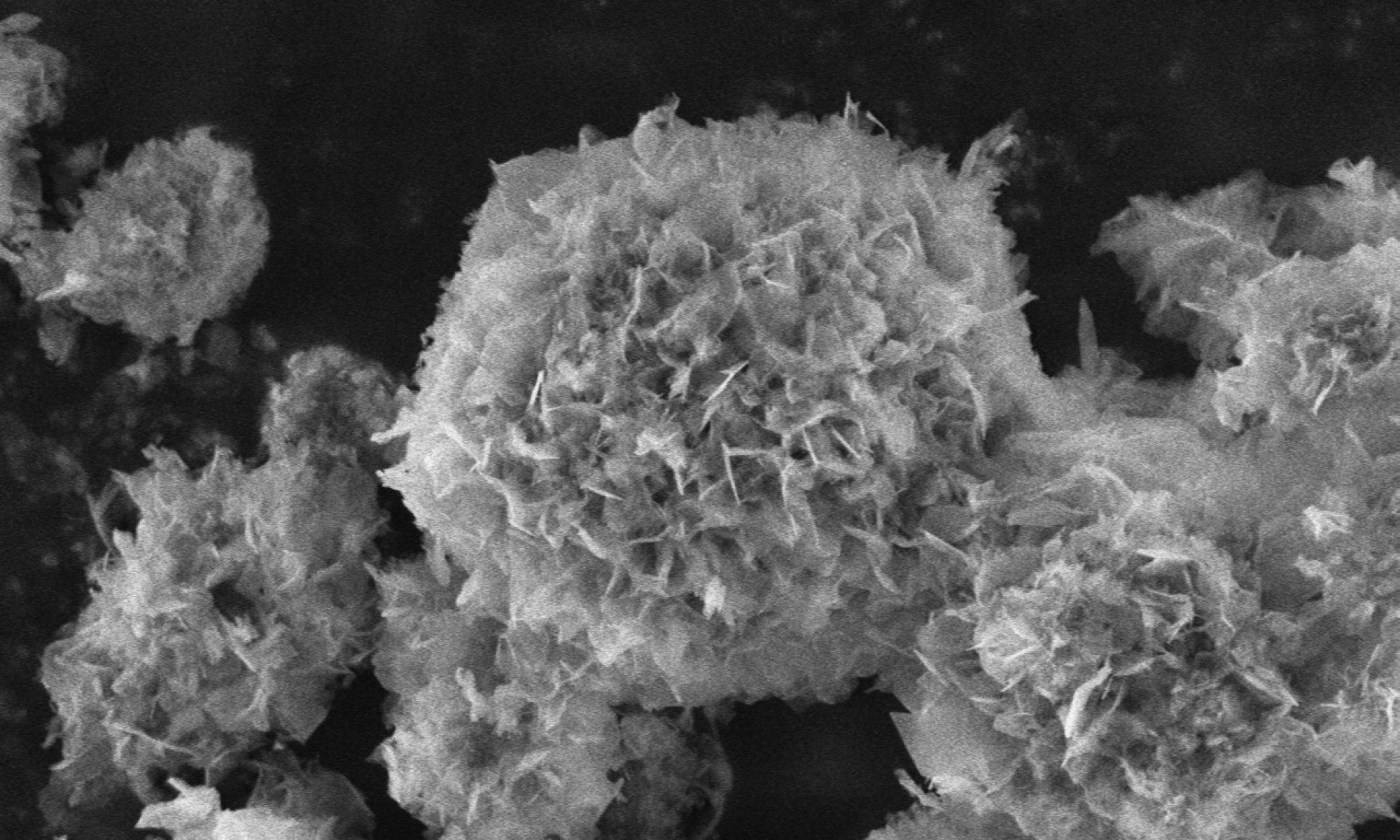
morphologies and their composites with calcium deficient hydroxyapatite (CaDHA/TiNMs) on hemostasis, as well as the cell viability and inflammatory response of Jurkat T cells. Four different TiNMs, nanoparticles (TiNPs), nanoplates (TiNPls), nanotubes (TiNTs), nanowires (TiNWs), and their composites with CaDHA were studied.
Among investigated materials, the least effect was observed for CaDHA. Effect on plasma coagulation was observed only for TiNPs, which shortened the prothrombin time. All TiNMs affected global hemostasis, especially the elongated TiNWs. Coating TiNMs with CaDHA resulted in a less pronounced effect compared to TiNMs. No significant effect on the viability of Jurkat T cells was observed for TiNMs, while composites increased it. Analysis of the expression of pro- inflammatory (IL-6, IL-8 and TNFa) and anti-inflammatory (IL-10) cytokines indicated inflammatory potential of investigated materials. Among TiNMs, TiNWs had the most pronounced pro-inflammatory effect, while CaDHA/TiNTs and CaDHA/TiNWs showed the most pronounced effect among composites.  
The obtained results confirm the potential of investigated materials for biomedical
applications. However, further studies are needed to establish a more precise relationship between the materials’ physico-chemical properties and their biological effects, thus ensuring their safe application.

Paper titled “Comparison of bovine serum albumin and chitosan effect on calcium phosphate formation in the presence of silver nanoparticles” is accepted for publishing in RSC Advances

The paper is authored by: Suzana Inkret, Ina Erceg, Marija Ćurlin, Nikolina Kalčec, Nikolina Peranić, Ivana Vinković Vrček, Darija Domazet Jurašin and Maja Dutour Sikirić

The article is available here:
Comparison of bovine serum albumin and chitosan effects on calcium phosphate formation in the presence of silver nanoparticles – RSC Advances (RSC Publishing)

Abstract:
The precipitation of calcium phosphates (CaPs) in the presence of more than one type of additive is of interest both from a fundamental point of view and as a possible biomimetic route for the preparation of multicomponent composites in which the activity of the components is preserved. In this study, the effect of bovine serum albumin (BSA) and chitosan (Chi) on the precipitation of CaPs in the presence of silver nanoparticles (AgNPs) stabilized with sodium bis(2-ethylhexyl)sulfosuccinate (AOT-AgNPs), poly(vinylpyrrolidone) (PVP-AgNPs), and citrate (cit-AgNPs) was investigated. In the control system, the precipitation of CaPs occurred in two steps. Amorphous calcium phosphate (ACP) was the first precipitated solid, which transformed into a mixture of calcium-deficient hydroxyapatite (CaDHA) and a smaller amount of octacalcium phosphate (OCP) after 60 min of ageing. Both biomacromolecules inhibited ACP transformation, with Chi being a stronger inhibitor due to its flexible molecular structure. As the concentration of the biomacromolecules increased, the amount of OCP decreased both in the absence and presence of AgNPs. In the presence of cit-AgNPs and two highest BSA concentrations, a change in the composition of the crystalline phase was observed. Calcium hydrogen phosphate dihydrate was formed in the mixture with CaDHA. An effect on the morphology of both the amorphous and crystalline phases was observed. The effect depended on the specific combination of biomacromolecules and differently stabilized AgNP. The results obtained suggest a simple method for fine-tuning the properties of precipitates using different classes of additives. This could be of interest for the biomimetic preparation of multifunctional composites for bone tissue engineering.